
Comprehensive Guide to Diagnosis and Management of Common Bile Duct Stones
- DR Dinesh Vats
- Aug 14, 2025
- 4 min read

The Common Bile Duct (CBD) is a channel that carries bile from the liver and gallbladder (GB) to the duodenum. The CBD opens into the second part of the duodenum at the Ampulla of Vater. Stones in the CBD may originate from the gallbladder or may form primarily within the CBD itself.
The risk factors for CBD stones are the same as those for gallbladder stones.
Symptoms
Severe epigastric pain or right upper quadrant (RUQ) abdominal pain radiating to the back.
Pain is often aggravated after meals, especially heavy or fatty meals, and may be associated with vomiting.
In most patients, the pain does not have the typical radiation and presents only as severe epigastric pain.
In cases where the CBD becomes infected (cholangitis), patients may present with jaundice, fever, and chills.
When to Suspect CBD Stones
Severe epigastric or RUQ pain without other features of dyspepsia.
A patient with known gallbladder stones presenting with severe epigastric pain.
Development of jaundice or fever in a known case of gallbladder stones.
Elevated bilirubin or alkaline phosphatase (ALP) in a patient with gallbladder stones.
Diagnosis
1. Ultrasonography (USG) Abdomen
First-line investigation to screen the CBD.
The lower end of the CBD is often difficult to visualize due to bowel gas, reducing sensitivity and specificity.
In the absence of gallbladder stones and with normal liver function tests (LFTs), a normal CBD on USG does not require further investigation for CBD stones—alternative causes of pain should be explored.
In the presence of gallbladder stones and elevated ALP, further tests such as MRCP or Endoscopic Ultrasound (EUS) are required, even if the CBD appears normal on USG. (See Image. 1 for diagnostic guidelines.)
2. Magnetic Resonance Cholangiopancreatography (MRCP)
A non-invasive MRI-based imaging technique with high sensitivity and specificity for CBD stones.
3. Endoscopic Ultrasound (EUS)
Combines endoscopy and ultrasound for high-resolution imaging.
Superior to MRCP for detecting stones smaller than 5 mm.
Allows for ERCP to be performed in the same session under the same anesthesia, saving time and improving effici
ency.
In our center, EUS is preferred over MRCP as it facilitates same-session ERCP or cholecystectomy. (See Image 2.)

Image 1. Diagnostic alogrithm for suspected common bile duct stones(CBDSs). LFT, liver function tests;US;ultrasound;CBD, common bile duct;EUS, endoscopic ultrasonography;MRCP, magnetic resonance cholangiopancreatography;ERCP, endoscopic reterograde cholangiopancreatography. 
Protocol followed in our EUS Gastroenterology centre for diagnosis and management of CBD Stones. 
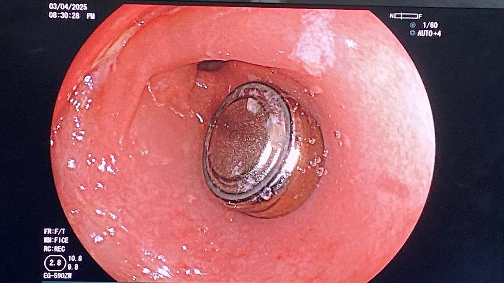
Image 2. Endoscopic ultrasound (EUS) image showing a common bile duct (CBD) stone, indicated by an arrow. Image credit: Dr. Naveen Kumar. Management
There is no medical (drug) treatment for CBD stones.
Stones can be removed surgically (laparoscopic or open CBD exploration) or via a minimally invasive endoscopic procedure known as Endoscopic Retrograde Cholangiopancreatography (ERCP).
ERCP is preferred over surgery in many cases because it is minimally invasive, usually performed as a day-care procedure under conscious sedation, and carries fewer complications.
In cases of cholangitis and stones smaller than 15 mm, ERCP is the procedure of choice.
Stones larger than 15 mm may require surgery or specialized ERCP techniques (to be discussed in the next article).
Case Summary
A 26-year-old female presented to our emergency department with severe epigastric pain radiating to the back for 3 days, aggravated by food intake and associated with vomiting. She also reported fever and jaundice for the past day. She had delivered a baby one month earlier and had gained approximately 10 kg during pregnancy.
Clinical suspicion: Cholangitis.
Examination findings: Icterus, fever, RUQ tenderness, and tachycardia.
Investigations:
USG abdomen showed multiple gallbladder stones (5 mm) and a dilated CBD containing a few small calculi (5 mm).
Given the diagnosis of cholangitis, she underwent emergency ERCP.
ERCP Procedure
Under sedation, a special side-viewing endoscope is inserted through the mouth into the duodenum.
The papilla is identified, and the CBD is cannulated under fluoroscopic guidance.

Image 3. Endoscopic view showing an impacted stone at the papilla in a patient with cholangitis. Image credit: Dr. Naveen Kumar. Contrast is injected into the CBD; under fluoroscopy, the CBD fills with contrast, and stones appear as filling defects.
Sphincterotomy is performed, and balloon dilatation of the papilla is done if needed.
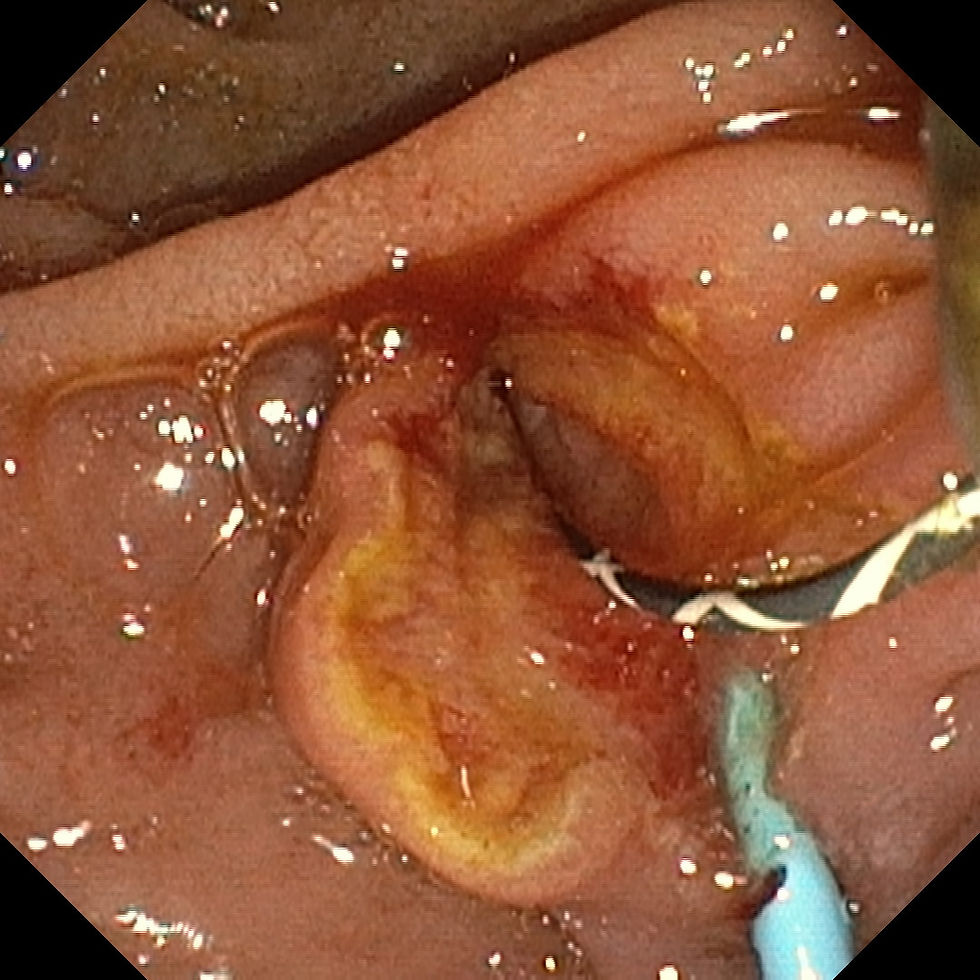
Image 4. Endoscopic view showing sphincterotomy performed prior to stone extraction. Image credit: Dr. Naveen Kumar. Balloon sweeps are used to remove stones into the duodenum.

Image 5. Endoscopic view showing balloon sweeps being performed to remove common bile duct (CBD) stones. Image credit: Dr. Naveen Kumar. Contrast is reinjected to confirm complete clearance of the CBD.

Image 6. Endoscopic view showing multiple stones in the duodenum after removal via balloon sweeps. Image credit: Dr. Naveen Kumar. A plastic stent is placed in the CBD to ensure free bile drainage into the duodenum.
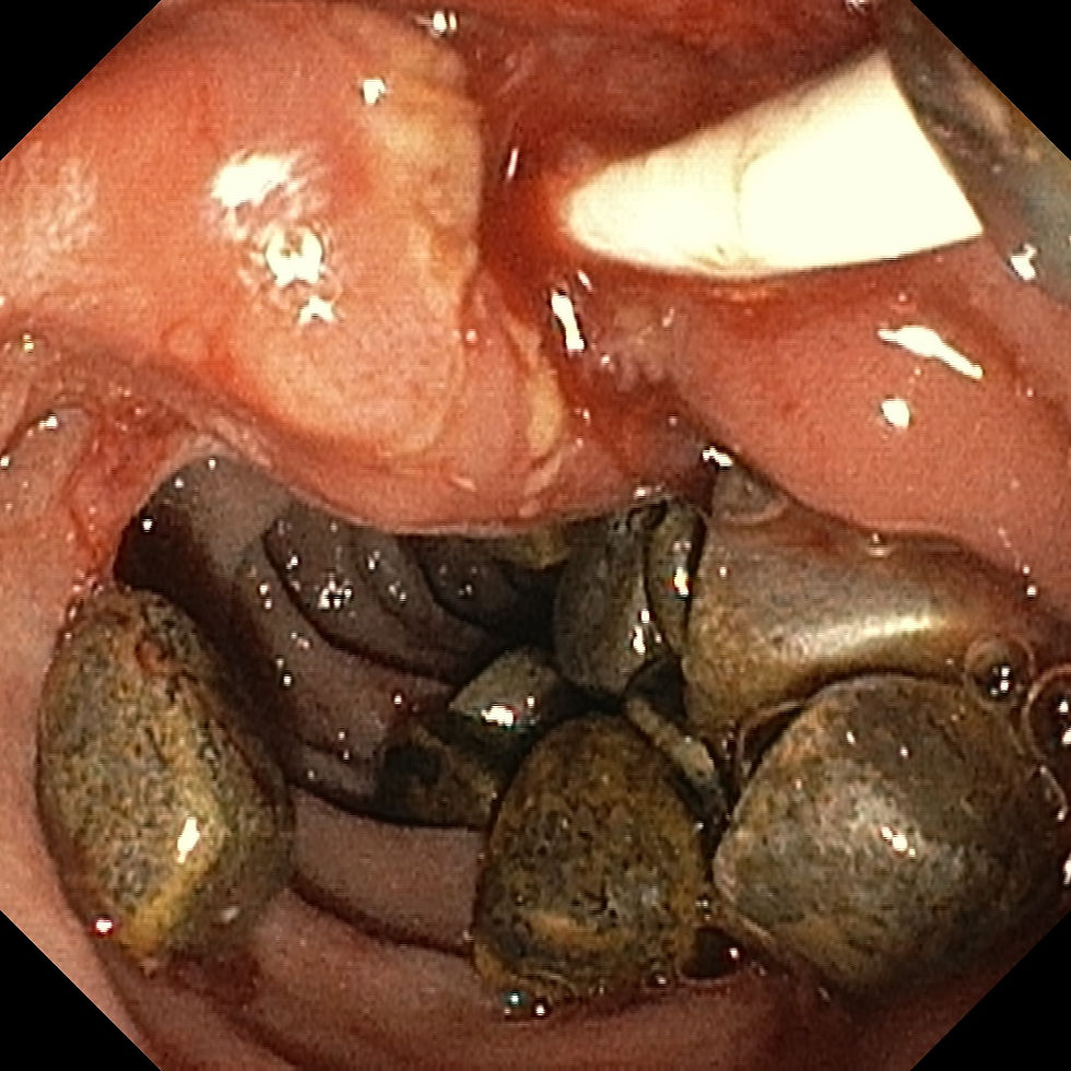
Image 7. Endoscopic image showing multiple common bile duct (CBD) stones being removed during balloon sweeps. Image credit: Dr. Naveen Kumar. 
Image 8. Endoscopic image showing multiple common bile duct (CBD) stones alongside biliary stents during balloon sweep removal. Image credit: Dr. Naveen Kumar. 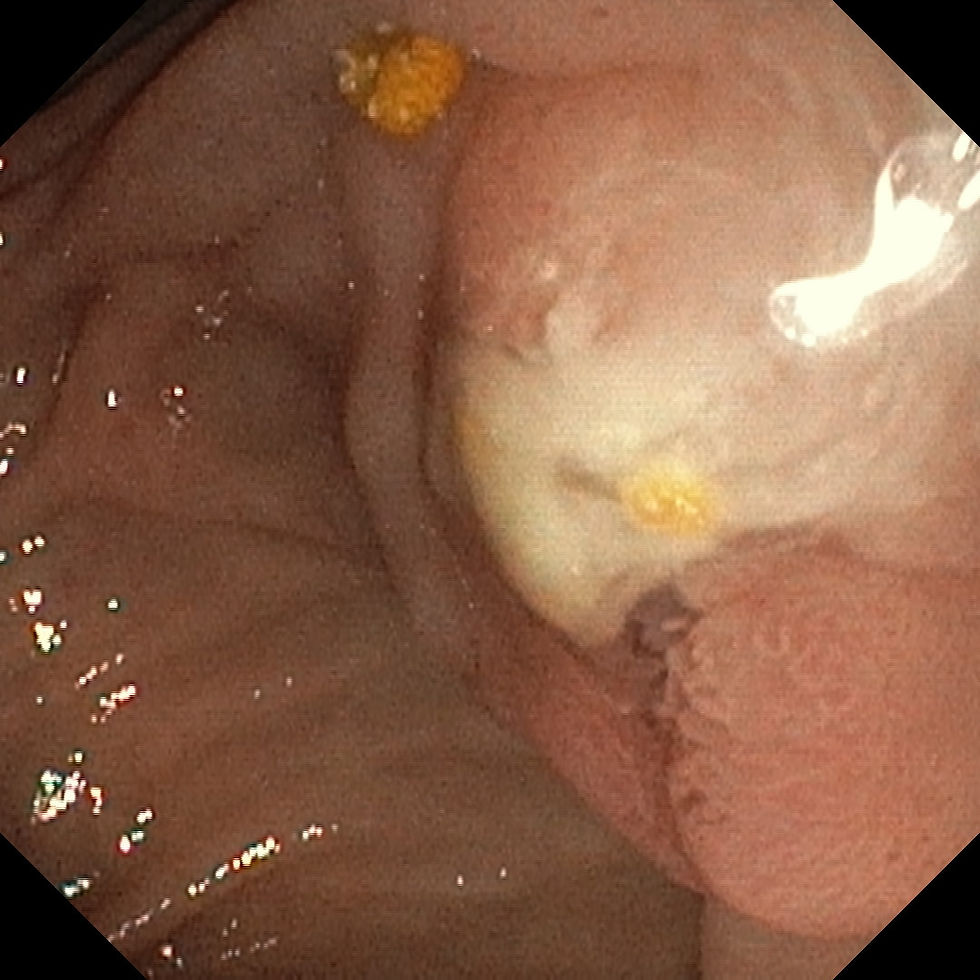
Image 9. Endoscopic image showing pus discharge from the papilla in a patient with severe cholangitis. Image credit: Dr. Naveen Kumar.
Conclusion
Common Bile Duct stones are a potentially serious condition that can lead to life-threatening infections like cholangitis.Early recognition based on history, clinical presentation, and targeted imaging is crucial.ERCP remains the treatment of choice in most cases due to its minimally invasive nature, quick recovery, and high success rate.For best outcomes, a epwise diagnostic approach and timely intervention are essential.
About the Author
Dr. Naveen Kumar, MBBS, MD, DM (Gastroenterology, PGIMER Chandigarh)Director, Department of Gastroenterology Sri Harihar Hospital and Research Center, Gutkar, District Mandi, Himachal Pradesh.

Dr. Naveen Kumar
Dr. Naveen Kumar is a highly respected gastroenterologist with advanced training from the prestigious PGIMER, Chandigarh. As the Director of the Department of Gastroenterology at Sri Harihar Hospital and Research Center, he brings exceptional expertise in diagnosing and managing a wide spectrum of digestive disorders, with a strong focus on advanced endoscopic techniques.
He is the first and only gastroenterologist in Himachal Pradesh performing endoscopic ultrasound (EUS) and cutting-edge procedures like Peroral Endoscopic Myotomy (POEM) for achalasia cardia, setting new standards in gastrointestinal care across the region.
Renowned for his patient-centered care and commitment to clinical excellence, Dr. Naveen also plays an active role in medical education and public awareness through his writings and academic contributions.
He is the editor of this article, and all endoscopic images included are credited to him.
📧 Email: contact@drnaveenkumargastro.com🐦 Twitter: @drnaveenm

Comments